23+ Hbme Immunostain Images. The term immunostaining was originally used to refer to the immunohistochemical staining of tissue sections, as first described by albert coons in 1941. When positive it is suggestive of malignancy, but when negative, it is not helpful.
This process uses an antibody targeted against a specific molecule.
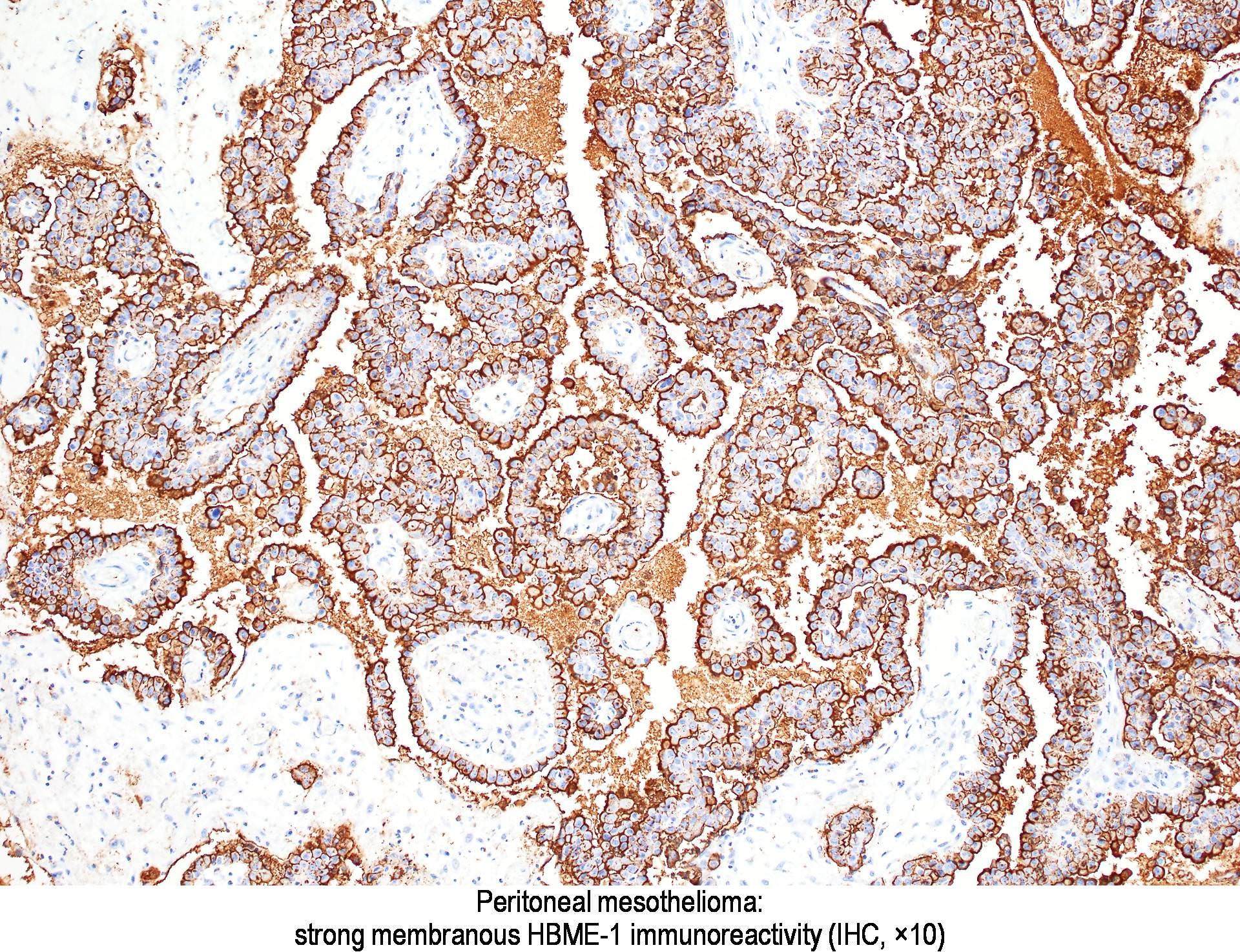
Aliases lists additional common names for a test, as an aid in searching. Xu b, ghossein r, lane j, lin o, katabi n. It stains normal mesothelial cells as well as epithelial mesotheliomas. When positive it is suggestive of malignancy, but when negative, it is not helpful.
